
Grad Life: What’s an MRI without the I?
-
-
slice.mit.edu
Filed Under


I remember sitting in 8th grade history class watching a documentary about the Ford Model T. A woman on the screen recalled the first time she sat behind the wheel of the iconic vehicle. “I felt so powerful controlling that big machine!” she told the camera.
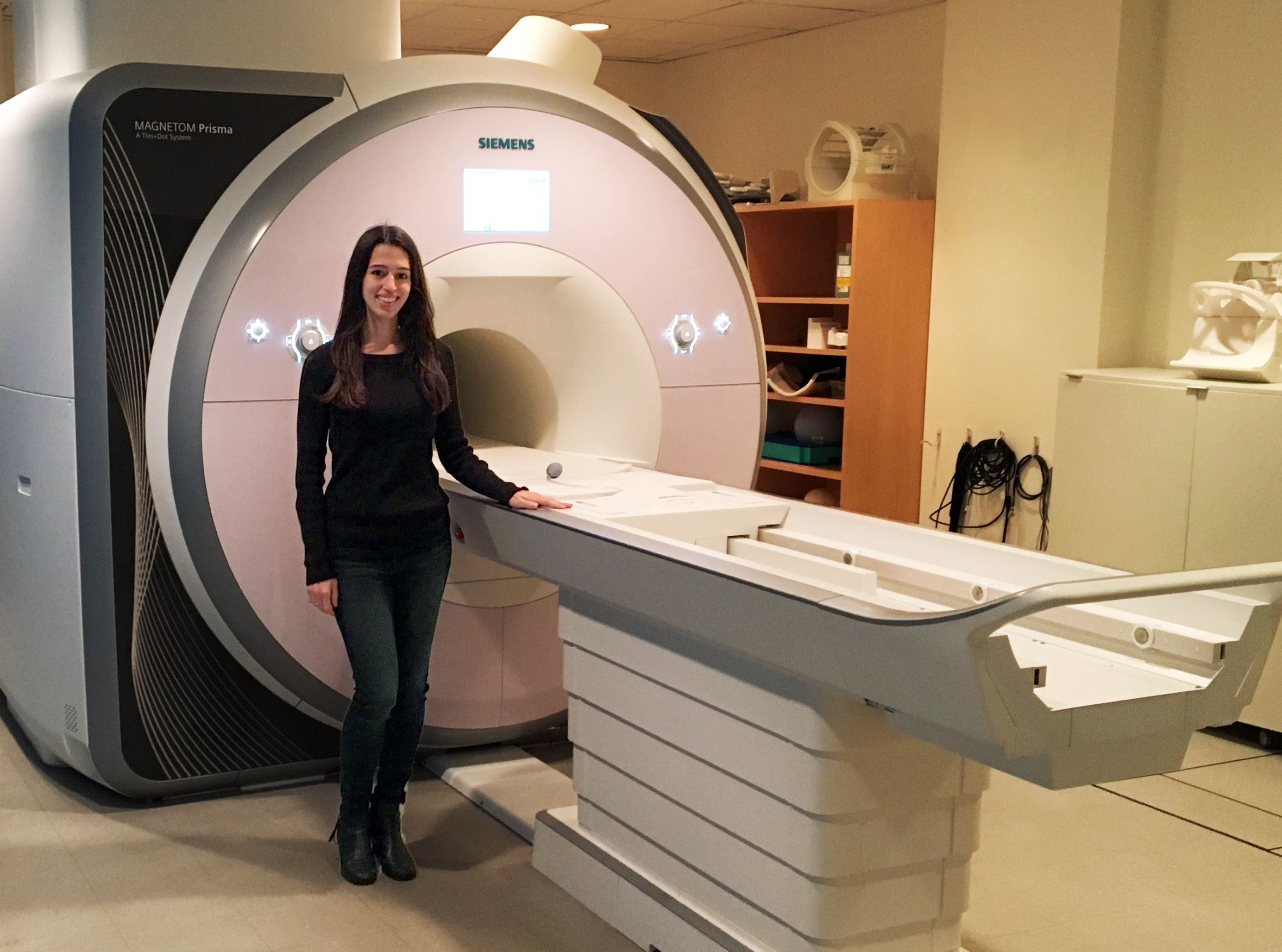
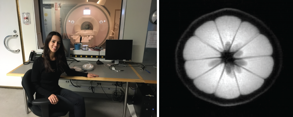
The documentary came back to my mind the first time I sat behind the controls of a magnetic resonance imaging (MRI) scanner: I finally felt exactly like that woman in the documentary.
I pressed the “scan” button of the $2 million machine. Whirrrr whirrrr whirrrr. Two minutes later I was looking at the inside of a lemon. I had never felt so cool in my entire life.
Magnetic resonance is the most beautiful physical phenomenon humankind has been able to harness. Research in magnetic resonance has produced more Nobel Prizes than any other area. Its utility in medicine is unmatched. And its elegant mathematics have captivated physicists, engineers, and computer scientists for decades.
Unlike its CT, PET, and X-ray cousins, MRI releases no ionizing radiation. You can have thousands of MRI scans in your life and never increase your chances of developing cancer. Even better, MRI scans permit doctors to see things inside the living body that are not possible to see in any other way.
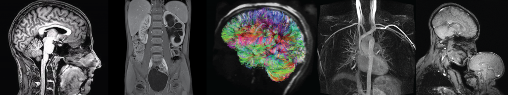
All the beautiful MRI images you see in Figure 2 come back to Faraday’s Law of Induction. Perhaps you’ve done an experiment in science class where you insert a magnet inside a copper coil. When you do this, a voltage is induced in the coil. In a similar way, MRI images are generated by atoms in your body inducing a voltage in a coil. Watch this five-minute video to understand how it works.
My PhD explores the topic: is it possible to obtain similar diagnostic information as we get from MRI but at much lower cost? It turns out, yes! When we remove the “I” (imaging) from MRI, the system gets a lot simpler and cheaper.
To generate images, you need incredibly precise magnetic gradient fields, extreme homogeneity of the main magnetic field, and experienced (note: expensive) doctors to interpret the images. All of these factors add up to make MRI one of the most expensive tests that a doctor can order.
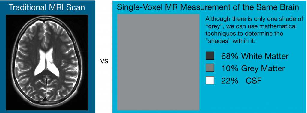
When you don’t generate images, it means you treat your entire measurement sample as one pixel. Now, instead of getting images of the brain where each pixel is a different shade of grey, we just have one pixel and one shade of gray.
Even though we only have one shade of gray, we can use different mathematical techniques to figure out the individual “shades” that it’s composed of. We can get nearly the same information (except for location info) as with a quantitative MRI scan, but without the need to generate an image. The magnetic resonance (MR) sensor is cheaper, smaller, and faster because of it.
My research applies this same technique to the human body. I am developing a non-invasive hydration sensor using magnetic resonance. There are many diseases—like congestive heart failure and chronic kidney disease—where the body accumulates too much fluid and people essentially drown in their own body water. There are no accurate, non-invasive, and quantitative ways to measure this fluid accumulation today. Cheaper, single-voxel MR sensors are perfect for the job.
Our lab builds custom MR sensors that you can hold up to different parts of the body. We use those sensors to collect data on fluid-overloaded patients at MGH, and then we develop algorithms to make sense of that data and relate it to fluid accumulation.
Hopefully one day you’ll go in for a regular checkup and your doctor will measure your hydration level with a portable MR sensor just like they measure your blood pressure with a sphygmomanometer. And hopefully, that will be just the beginning of an era of medicine where we use the power of magnetic resonance not just in multi-million dollar MRI suites but in the doctor’s office, pharmacy, and even the home.
Grad Life blog posts offer insights from current MIT graduate students on Slice of MIT.


