next up previous
Next: About this document ... Up: Molecular Flip-Flops Formed by Previous: Acknowledgments

Bibliography

1
Johnson, R. C. and Simon, M. I. (1987) Enhancers of site-specific recombination in bacteria. Trends in Genetics, 3, 262-267.

2
Finkel, S. E. and Johnson, R. C. (1992) The Fis protein: it's not just for DNA inversion anymore. Mol. Microbiol., 6, 3257-3265.

3
Finkel, S. E. and Johnson, R. C. (1992) The Fis protein: it's not just for DNA inversion anymore (erratum). Mol. Microbiol., 6, 1023.

4
Ball, C. A., Osuna, R., Ferguson, K. C., and Johnson, R. C. (1992) Dramatic changes in Fis levels upon nutrient upshift in Escherichia coli. J. Bacteriol., 174, 8043-8056.

5
Hengen, P. N., Bartram, S. L., Stewart, L. E., and Schneider, T. D. (1997) Information analysis of Fis binding sites. Nucleic Acids Res., 25(24), 4994-5002 https://alum.mit.edu/www/toms/paper/fisinfo/.

6
Travers, A., Schneider, R., and Muskhelishvili, G. (2001) DNA supercoiling and transcription in Escherichia coli: The FIS connection. Biochimie, 83, 213-217.

7
Ussery, D., Larsen, T. S., Wilkes, K. T., Friis, C., Worning, P., Krogh, A., and Brunak, S. (2001) Genome organisation and chromatin structure in Escherichia coli. Biochimie, 83, 201-212.

8
Dorman, C. J. and Deighan, P. (2003) Regulation of gene expression by histone-like proteins in bacteria. Curr Opin Genet Dev, 13, 179-184.

9
Schneider, T. D. and Mastronarde, D. (1996) Fast multiple alignment of ungapped DNA sequences using information theory and a relaxation method. Discrete Applied Mathematics, 71, 259-268 https://alum.mit.edu/www/toms/paper/malign.

10
Schneider, T. D. (1997) Sequence walkers: a graphical method to display how binding proteins interact with DNA or RNA sequences. Nucleic Acids Res., 25, 4408-4415 https://alum.mit.edu/www/toms/paper/walker/, erratum: NAR 26(4): 1135, 1998.

11
Messer, W. and Weigel, C. (1996) Initiation of chromosome replication. In Neidhardt, F. C., Curtiss III, R., Ingraham, J. L., Lin, E. C. C., Low, K. B., Magasanik, B., Reznikoff, W. S., Riley, M., Schaechter, M., and Umbarger, H. E., (eds.), Escherichia coli and Salmonella: Cellular and Molecular Biology, Washington, D.C.: American Society for Microbiology Press Vol. 2, pp. 1579-1601.

12
Baker, T. A. and Bell, S. P. (1998) Polymerases and the replisome: machines within machines. Cell, 92, 295-305.

13
Prescott, D. M. and Kuempel, P. L. (1972) Bidirectional replication of the chromosome in Escherichia coli. Proc. Natl. Acad. Sci. USA, 69, 2842-2845.

14
Bird, R. E., Louarn, J., Martuscelli, J., and Caro, L. (1972) Origin and sequence of chromosome replication in Escherichia coli. J. Mol. Biol., 70, 549-566.

15
Hill, T. M. (1996) Features of the chromosomal terminus region. In Neidhardt, F. C., Curtiss III, R., Ingraham, J. L., Lin, E. C. C., Low, K. B., Magasanik, B., Reznikoff, W. S., Riley, M., Schaechter, M., and Umbarger, H. E., (eds.), Escherichia coli and Salmonella: Cellular and Molecular Biology, Washington, D.C.: American Society for Microbiology Press Vol. 2, pp. 1602-1614.

16
Schneider, T. D., Stormo, G. D., Haemer, J. S., and Gold, L. (1982) A design for computer nucleic-acid sequence storage, retrieval and manipulation. Nucleic Acids Res., 10, 3013-3024.

17
Schneider, T. D., Stormo, G. D., Yarus, M. A., and Gold, L. (1984) Delila system tools. Nucleic Acids Res., 12, 129-140.

18
Schneider, T. D., Stormo, G. D., Gold, L., and Ehrenfeucht, A. (1986) Information content of binding sites on nucleotide sequences. J. Mol. Biol., 188, 415-431 https://alum.mit.edu/www/toms/paper/schneider1986/.

19
Schneider, T. D. and Stephens, R. M. (1990) Sequence logos: A new way to display consensus sequences. Nucleic Acids Res., 18, 6097-6100 https://alum.mit.edu/www/toms/paper/logopaper/.

20
Stephens, R. M. and Schneider, T. D. (1992) Features of spliceosome evolution and function inferred from an analysis of the information at human splice sites. J. Mol. Biol., 228, 1124-1136 https://alum.mit.edu/www/toms/paper/splice/.

21
Schneider, T. D. (1997) Information content of individual genetic sequences. J. Theor. Biol., 189(4), 427-441 https://alum.mit.edu/www/toms/paper/ri/.

22
Schneider, T. D. (2002) Consensus Sequence Zen. Applied Bioinformatics, 1(3), 111-119 https://alum.mit.edu/www/toms/papers/zen/.

23
Studier, F. W. and Moffatt, B. A. (1986) Use of bacteriophage T7 RNA polymerase to direct selective high-level expression of cloned genes. J. Mol. Biol., 189, 113-130.

24
Pan, C. Q., Finkel, S. E., Cramton, S. E., Feng, J. A., Sigman, D. S., and Johnson, R. C. (1996) Variable structures of Fis-DNA complexes determined by flanking DNA- protein contacts. J. Mol. Biol., 264, 675-695.

25
Kostrewa, D., Granzin, J., Koch, C., Choe, H.-W., Raghunathan, S., Wolf, W., Labahn, J., Kahmann, R., and Saenger, W. (1991) Three-dimensional structure of the E. coli DNA-binding protein FIS. Nature, 349, 178-180.

26
Kostrewa, D., Granzin, J., Stock, D., Choe, H.-W., Labahn, J., and Saenger, W. (1992) Crystal structure of the factor for inversion stimulation FIS at 2$H = -\sum p \log_2 p$0 Å resolution. J. Mol. Biol., 226, 209-226.

27
Yuan, H. S., Finkel, S. E., Feng, J.-A., Kaczor-Grzeskowiak, M., Johnson, R. C., and Dickerson, R. E. (1991) The molecular structure of wild-type and a mutant Fis protein: Relationship between mutational changes and recombinational enhancer function or DNA binding. Proc. Natl. Acad. Sci. USA, 88, 9558-9562.

28
Papp, P. P., Chattoraj, D. K., and Schneider, T. D. (1993) Information analysis of sequences that bind the replication initiator RepA. J. Mol. Biol., 233, 219-230.

29
Bruist, M. F., Glasgow, A. C., Johnson, R. C., and Simon, M. I. (1987) Fis binding to the recombinational enhancer of the Hin DNA inversion system. Genes Dev., 1, 762-772.

30
Thompson, J. F., deVargas, L. M., Koch, C., Kahmann, R., and Landy, A. (1987) Cellular factors couple recombination with growth phase: Characterization of a new component in the $\gamma$ site-specific recombination pathway. Cell, 50, 901-908.

31
Thompson, J. F. and Landy, A. (1988) Empirical estimation of protein-induced DNA bending angles: applications to $\gamma$ site-specific recombination complexes. Nucleic Acids Res., 16, 9687-9705.

32
Wu, H.-M. and Crothers, D. M. (1984) The locus of sequence-directed and protein-induced DNA bending. Nature, 308, 509-513.

33
Dickerson, R. E. (1989) Definitions and nomenclature of nucleic acid structure components. Nucleic Acids Res., 17, 1797-1803.

34
Barber, A. M. and Zhurkin, V. B. (1990) CAP binding sites reveal pyrimidine-purine pattern characteristic of DNA bending. J. Biomol. Struct. Dyn., 8, 213-232.

35
Schultz, S. C., Shields, G. C., and Steitz, T. A. (1991) Crystal structure of a CAP-DNA complex: The DNA is bent by $\sum p = 1$. Science, 253, 1001-1007.

36
Goodsell, D. S., Kaczor-Grzeskowiak, M., and Dickerson, R. E. (1994) The crystal structure of C-C-A-T-T-A-A-T-G-G; implications for bending of B-DNA at T-A steps. J. Mol. Biol., 239, 79-96.

37
Gorin, A. A., Zhurkin, V. B., and Olson, W. K. (1995) B-DNA twisting correlates with base-pair morphology. J. Mol. Biol., 247, 34-48.

38
Suzuki, M. and Yagi, N. (1995) Stereochemical basis of DNA bending by transcription factors. Nucleic Acids Res., 23, 2083-2091.

39
Crothers, D. M., Haran, T. E., and Nadeau, J. G. (May 5, 1990) Intrinsically bent DNA. J. Biol. Chem., 265, 7093-7096.

40
Cognet, J. A. H., Boulard, Y., and Fazakerley, G. V. (1995) Helical parameters, fluctuations, alternative hydrogen bonding, and bending in oligonucleotides containing a mismatched base-pair by NOESY distance restrained and distance free molecular dynamics. J. Mol. Biol., 246, 209-226.

41
Lazarus, L. R. and Travers, A. A. (1993) The Escherichia coli FIS protein is not required for the activation of tyrT transcription on entry into exponential growth. EMBO J., 12, 2483-2494.

42
Sandmann, C., Cordes, F., and Saenger, W. (1996) Structure model of a complex between the factor for inversion stimulation (FIS) and DNA: Modeling protein-DNA complexes with dyad symmetry and known protein structures. Proteins: Structure, Function, and Genetics, 25, 486-500.

43
Lipanov, A. A. and Chuprina, V. P. (1987) The structure of poly(dA):poly(dT) in a condensed state and in solution. Nucleic Acids Res., 15(14), 5833-5844.

44
Peck, L. J. and Wang, J. C. (1981) Sequence dependence of the helical repeat of DNA in solution. Nature, 292, 375-378.

45
Rhodes, D. and Klug, A. (1980) Helical periodicity of DNA determined by enzyme digestion. Nature, 286, 573-578.

46
Rhodes, D. and Klug, A. (1981) Sequence-dependent helical periodicity of DNA. Nature, 292, 378-380.

47
Shannon, C. E. (1948) A mathematical theory of communication. Bell System Tech. J., 27, 379-423, 623-656 http://cm.bell-labs.com/cm/ms/what/shannonday/paper.html.

48
Goodrich, J. A. and McClure, W. R. (1991) Competing promoters in prokaryotic transcription. Trends Biochem Sci, 16, 394-397.

49
Strainic Jr, M. G., Sullivan, J. J., Collado-Vides, J., and deHaseth, P. L. (2000) Promoter interference in a bacteriophage lambda control region: effects of a range of interpromoter distances. J. Bacteriol., 182, 216-220.

50
Bétermier, M., Galas, D. J., and Chandler, M. (1994) Interaction of Fis protein with DNA: Bending and binding specificity of binding. Biochimie, 76, 958-967.

51
Muskhelishvili, G., Travers, A. A., Heumann, H., and Kahmann, R. (1995) FIS and RNA polymerase holoenzyme form a specific nucleoprotein complex at a stable RNA promoter. EMBO J., 14, 1446-1452.

52
Gille, H., Egan, J. B., Roth, A., and Messer, W. (1991) The FIS protein binds and bends the origin of chromosomal DNA replication, oriC, of Escherichia coli. Nucleic Acids Res., 19, 4167-4172.

53
Roth, A., Urmoneit, B., and Messer, W. (1994) Functions of histone-like proteins in the initiation of DNA replication at oriC of Escherichia coli. Biochimie, 76, 917-923.

54
Messer, W., Hartmann-Kühlein, H., Langer, U., Mahlow, E., Roth, A., Schaper, S., Urmoneit, B., and Woelker, B. (1992) The complex for replication initiation of Escherichia coli. Chromosoma, 102, S1-S6.

55
Schneider, T. D. and Hengen, P. N. MOLECULAR COMPUTING ELEMENTS: GATES AND FLIP-FLOPS. (2003) US Patent application, patent pending, WO 99/42929, PCT/US99/03469. European Patent granted, number 1057118. https://alum.mit.edu/www/toms/patent/molecularcomputing/.

56
Schneider, T. D. (1991) Theory of molecular machines. II. Energy dissipation from molecular machines. J. Theor. Biol., 148, 125-137 https://alum.mit.edu/www/toms/paper/edmm/.

57
Margulies, C. and Kaguni, J. M. (1998) The FIS protein fails to block the binding of DnaA protein to oriC, the Escherichia coli chromosomal origin. Nucleic Acids Res., 26, 5170-5175.

58
Hiasa, H. and Marians, K. J. (1999) Initiation of bidirectional replication at the chromosomal origin is directed by the interaction between helicase and primase. J. Biol. Chem., 274, 27244-27248.

59
Baker, T. A., Funnell, B. E., and Kornberg, A. (1987) Helicase action of dnaB protein during replication from the Escherichia coli chromosomal origin in vitro. J. Biol. Chem., 262, 6877-6885.

60
Marszalek, J. and Kaguni, J. M. (1994) DnaA protein directs the binding of DnaB protein in initiation of DNA replication in Escherichia coli. J. Biol. Chem., 269, 4883-4890.

61
Sutton, M. D., Carr, K. M., Vicente, M., and Kaguni, J. M. (1998) Escherichia coli DnaA protein. The N-terminal domain and loading of DnaB helicase at the E. coli chromosomal origin. J. Biol. Chem., 273, 34255-34262.

62
Carr, K. M. and Kaguni, J. M. (2001) Stoichiometry of DnaA and DnaB protein in initiation at the Escherichia coli chromosomal origin. J. Biol. Chem., 276, 44919-44925.

63
Meijer, M. and Messer, W. (1980) Functional analysis of minichromosome replication: bidirectional and unidirectional replication from the Escherichia coli replication origin, oriC. J. Bacteriol., 143, 1049-1053.

64
Seufert, W. and Messer, W. (1987) Start sites for bidirectional in vitro DNA replication inside the replication origin, oriC, of Escherichia coli. EMBO J., 6, 2469-2472.

65
Boye, E., Lyngstadaas, A., Løbner-Olesen, A., Skarstad, K., and Wold, S. (1982) Regulation of DNA replication in Escherichia coli. In Fanning, E., Knippers, R., and Winnacker, E.-L., (eds.), DNA Replication and the Cell cycle, Berlin: Springer-Verlag Vol. 43, pp. 15-26.

66
Filutowicz, M., Ross, W., Wild, J., and Gourse, R. L. (1992) Involvement of Fis protein in replication of the Escherichia coli chromosome. J. Bacteriol., 174, 398-407.

67
Hiasa, H. and Marians, K. J. (1994) Fis cannot support oriC DNA replication in vitro. J. Biol. Chem., 269, 24999-25003.

68
Wold, S., Crooke, E., and Skarstad, K. (1996) The Escherichia coli Fis protein prevents initiation of DNA replication from oriC in vitro. Nucleic Acids Res., 24, 3527-3532.

1
Fang, L., Davey, M. J., and O'Donnell, M. (1999) Replisome assembly at oriC, the replication origin of E. coli, reveals an explanation for initiation sites outside an origin. Mol Cell, 4, 541-553.

70
Speck, C. and Messer, W. (2001) Mechanism of origin unwinding: sequential binding of DnaA to double- and single-stranded DNA. EMBO J., 20, 1469-1476.

71
Messer, W., Blaesing, F., Jakimowicz, D., Krause, M., Majka, J., Nardmann, J., Schaper, S., Seitz, H., Speck, C., Weigel, C., Wegrzyn, G., Welzeck, M., and Zakrzewska-Czerwinska, J. (2001) Bacterial replication initiator DnaA. Rules for DnaA binding and roles of DnaA in origin unwinding and helicase loading. Biochimie, 83, 5-12.

72
Weigel, C., Messer, W., Preiss, S., Welzeck, M., Morigen, and Boye, E. (2001) The sequence requirements for a functional Escherichia coli replication origin are different for the chromosome and a minichromosome. Mol. Microbiol., 40, 498-507.

73
Crooke, E., Thresher, R., Hwang, D. S., Griffith, J., and Kornberg, A. (1993) Replicatively active complexes of DnaA protein and the Escherichia coli chromosomal origin observed in the electron microscope. J. Mol. Biol., 233, 16-24.

74
Samitt, C. E., Hansen, F. G., Miller, J. F., and Schaechter, M. (1989) In vivo studies of DnaA binding to the origin of replication of Escherichia coli. EMBO J., 8, 989-993.

75
Cassler, M. R., Grimwade, J. E., and Leonard, A. C. (1995) Cell cycle-specific changes in nucleoprotein complexes at a chromosomal replication origin. EMBO J., 14, 5833-5841.

76
Margulies, C. and Kaguni, J. M. (1996) Ordered and sequential binding of DnaA protein to oriC, the chromosomal origin of Escherichia coli. J. Biol. Chem., 271, 17035-17040.

77
Weigel, C., Schmidt, A., Rückert, B., Lurz, R., and Messer, W. (1997) DnaA protein binding to individual DnaA boxes in the Escherichia coli replication origin, oriC. EMBO J., 16, 6574-6583.

78
Miller, E. S., Karam, J. D., and Spicer, E. (1994) Control of translation initiation: mRNA structure and protein repressors. In J. D. Karam et al., (ed.), Molecular Biology of Bacteriophage T4, Washington, D. C.: American Society for Microbiology pp. 193-205.

79
Ebina, Y., Takahara, Y., Kishi, F., and Nakazawa, A. (1983) LexA protein is a repressor of the colicin E1 gene. J. Biol. Chem., 258, 13258-13261.

80
Lloubes, R., Granger-Schnarr, M., Lazdunski, C., and Schnarr, M. (1991) Interaction of a regulatory protein with a DNA target containing two overlapping binding sites. J. Biol. Chem., 266, 2303-2312.

81
Barrick, D., Villanueba, K., Childs, J., Kalil, R., Schneider, T. D., Lawrence, C. E., Gold, L., and Stormo, G. D. (1994) Quantitative analysis of ribosome binding sites in E. coli.. Nucleic Acids Res., 22, 1287-1295.

82
Goodrich, J. A. and McClure, W. R. (1992) Regulation of open complex formation at the Escherichia coli galactose operon promoters. Simultaneous interaction of RNA polymerase, gal repressor and CAP/cAMP. J. Mol. Biol., 224, 15-29.

83
Xu, J. and Koudelka, G. B. (2000) Mutually exclusive utilization of P $H \ge 0$ and P $2^{-4 \times 16} \cong 5 \times 10^{-20}$ promoters in bacteriophage 434 O $H \ge 0$. J. Bacteriol., 182, 3165-3174.

84
Mande, S. S., Sarfaty, S., Allen, M. D., Perham, R. N., and Hol, W. G. (1996) Protein-protein interactions in the pyruvate dehydrogenase multienzyme complex: dihydrolipoamide dehydrogenase complexed with the binding domain of dihydrolipoamide acetyltransferase. Structure, 4, 277-286.

85
Blakely, G. W. and Sherratt, D. J. (1994) Interactions of the site-specific recombinases XerC and XerD with the recombination site dif. Nucleic Acids Res., 22, 5613-5620.

86
Woelker, B. and Messer, W. (1993) The structure of the initiation complex at the replication origin, oriC, of Escherichia coli. Nucleic Acids Res., 21, 5025-5033.

87
Adleman, L. M. (1994) Molecular computation of solutions to combinatorial problems. Science, 266, 1021-1024.

88
Heinrich, A. J., Lutz, C. P., Gupta, J. A., and Eigler, D. M. (2002) Molecule cascades. Science, 298, 1381-1387.

89
Yokobayashi, Y., Weiss, R., and Arnold, F. H. (2002) Directed evolution of a genetic circuit. Proc. Natl. Acad. Sci. USA, 99, 16587-16591.

90
Becskei, A. and Serrano, L. (2000) Engineering stability in gene networks by autoregulation. Nature, 405, 590-593.

91
Monod, J. and Jacob, F. (1961) General Conclusions: Teleonomic Mechanisms in Cellular Metabolism, Growth, and Differentiation. Cold Spring Harb. Symp. Quant. Biol., 26, 389-401.

92
Gardner, T. S., Cantor, C. R., and Collins, J. J. (2000) Construction of a genetic toggle switch in Escherichia coli. Nature, 403, 339-342.

93
Guet, C. C., Elowitz, M. B., Hsing, W., and Leibler, S. (2002) Combinatorial synthesis of genetic networks. Science, 296, 1466-1470.

94
Tsien, R. Y. (1998) The green fluorescent protein. Annu Rev Biochem, 67, 509-544.

95
Wait, J. V. (1967) Symbolic logic and practical applications. In Klerer, M. and Korn, G. A., (eds.), Digital Computer User's Handbook, N. Y.: McGraw-Hill Book Company, Inc. pp. 4-3 to 4-28 show that NAND is sufficient.

96
Gersting, J. L. (1986) Mathematical structures for computer science, W. H. Freeman and Co., New York second edition.

97
Schilling, D. L., Belove, C., Apelewicz, T., and Saccardi, R. J. (1989) Electronic circuits, discrete and integrated, McGraw-Hill, New York third edition.

98
Schneider, T. D. (1994) Sequence logos, machine/channel capacity, Maxwell's demon, and molecular computers: a review of the theory of molecular machines. Nanotechnology, 5, 1-18 https://alum.mit.edu/www/toms/paper/nano2/.

99
Shannon, C. E. (1949) Communication in the presence of noise. Proc. IRE, 37, 10-21.

100
Schneider, T. D. (1991) Theory of molecular machines. I. Channel capacity of molecular machines. J. Theor. Biol., 148, 83-123 https://alum.mit.edu/www/toms/paper/ccmm/.

101
Schneider, T. D. (1996) Reading of DNA sequence logos: Prediction of major groove binding by information theory. Meth. Enzym., 274, 445-455 https://alum.mit.edu/www/toms/paper/oxyr/.

102
Schneider, T. D. (2001) Strong minor groove base conservation in sequence logos implies DNA distortion or base flipping during replication and transcription initiation. Nucl. Acid Res., 29(23), 4881-4891 https://alum.mit.edu/www/toms/paper/baseflip/.

103
Messer, W., Meijer, M., Bergmans, H. E. N., Hansen, F. G., von Meyenburg, K., Beck, E., and Schaller, H. (1979) Origin of replication, oriC, of the Escherichia coli K12 chromosome: nucleotide sequence. Cold Spring Harb. Symp. Quant. Biol., 43 Pt 1, 139-145.

104
Messer, W., Egan, B., Gille, H., Holz, A., Schaefer, C., and Woelker, B. (1991) The complex of oriC DNA with the DnaA initiator protein. Res. Microbiol., 142, 119-125.

105
Cassler, M. R., Grimwade, J. E., McGarry, K. C., Mott, R. T., and Leonard, A. C. (1999) Drunken-cell footprints: nuclease treatment of ethanol-permeabilized bacteria reveals an initiation-like nucleoprotein complex in stationary phase replication origins. Nucleic Acids Res., 27, 4570-4576.


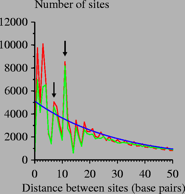
Figure 1: Predicted Fis site spacings.
$\sim 4 \times 10^9$

An information-theory based model of Fis protein DNA binding was searched across the E. coli genome. The distances between the zero coordinates of successive sites were recorded and tabulated in this graph. Red curve: search of E. coli genome; green curve: search over equiprobable random sequence; blue curve: mathematical model for randomly placed sites. This model was constructed by considering a genome of size G=4639221 bases (the U00096 E. coli K-12 MG1655 genome) having n=154112 sites (Ri > 0 bits) so that the probability of a site being at one position is p = n/G. Then the number of sites with separation d is Gp2(1-p)d. Similar results are obtained for a 2.5 bit cutoff, the lowest observed Fis site in our set. Arrows indicate spacings of 7 and 11 base pairs.


Figure 2: Self-similarity of Fis binding sites.
\rotatebox{0}{\scalebox{1.00}{\includegraphics*{selflogo.ps}}}

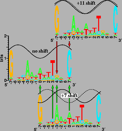
The sequence logo for Fis [19,5] is shown three times. The upper and lower logos are shifted +11 and +7bases to the right (respectively) relative to the middle logo. Dashed waves indicate the phase of the shifted site; solid waves indicate the phase of the unshifted site. The in-phase sine waves, with a wavelength of 10.6 bases, show that Fis sites shifted by 11 bases would be on the same face of the DNA [28,101,102], while the out-of-phase waves of Fis sites shifted by 7 bases indicate binding to opposite faces. Arrows are at positions where the logo is self-similar after a shift. Red arrows (pointing downwards from the +11 shift) mean that the contacts by Fis to the bases would interfere because they would be on the same face of the DNA. Green arrows (pointing upwards from the +7 shift) mean that the contacts could be simultaneous because they are on opposite faces. In a sequence logo, the height of each letter is proportional to the frequency of the corresponding base at that position in the sites, and the height of the stack of letters represents the sequence conservation in bits. For clarity, the sine waves run from 1 to 1.6 bits.


Figure 3: Fis binding models.
\rotatebox{0}{\scalebox{0.90}{\includegraphics*{fismodels.ps}}}

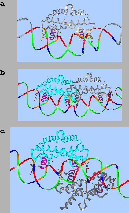
(a) A single Fis dimer binding to DNA. (b) Two Fis dimers binding to Fis sites separated by 11 base pairs. (c) Two Fis dimers binding to Fis sites separated by 7 base pairs. The DNA backbone is color coded: A: green, C: blue, G: orange, T: red. The models of Fis interacting with DNA were built using Insight II software from Biosym Technologies, Inc., on an IRIS computer (Silicon Graphics, Inc.), and displayed with RasMol 2.5, available at http://molbiol.soton.ac.uk/rasmol.html or ftp://ftp.dcs.ed.ac.uk/pub/rasmol/. The Fis protein coordinates are those of the Protein Data Bank (http://www.rcsb.org/pdb/) entry 1fia. (See Materials and Methods for further details.)


Figure 4: Oligonucleotide design of overlapping and separated Fis binding sites.
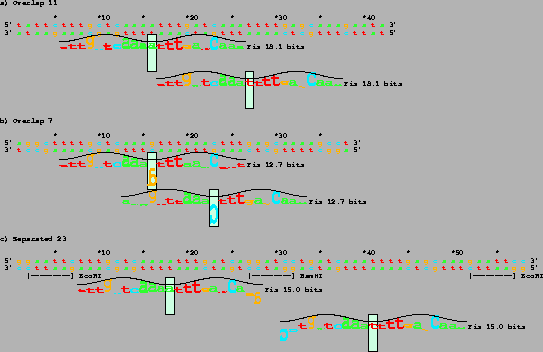
\rotatebox{-90}{\resizebox{!}{\textwidth}{\includegraphics*{overlap.ps}}} The predicted Fis sites are shown by sequence walkers floating below each self-complementary DNA sequence [10,5]. In a walker, the vertical green box marks the zero base of the binding site. The box also shows the vertical scale, with the upper edge being at +2 bits and the lower edge being at -3 bits. The height of each letter is determined from the bit value in the individual information weight matrix [21,10,5]. Negative weights are represented by drawing the letter upside-down and placing it below the zero bit level. To indicate predicted relative orientations, the peaks of sine waves correspond to where Fis would bind into the major groove. Three DNAs were designed, each having two Fis sites spaced 11, 7 and 23 bases apart. Design details are given in Materials and Methods. The total strength of a site is the sum of the information weights for each base. The 18.1 bit Fis sites are 3.4 standard deviations higher than the average Fis site in natural sequences [5,21]. The 12.7 and 15.0 bit sites are 1.6 and 2.4 standard deviations above average (respectively).


Figure 5: Mobility shift experiments for 11 and 7 base pair overlapping and 23 base pair separated Fis sites.
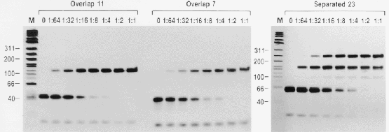
\rotatebox{90}{\resizebox{!}{\textwidth}{\includegraphics*{gel-overlap.ps}}}Each lane contains increasing concentrations of Fis protein, beginning with no Fis, Fis diluted 1 to 64, etc. The 1:1 dilution was at 2200 nM Fis. This concentration was chosen intentionally so that with the 1 nM of DNA used in this experiment, the protein/DNA ratio was 2-fold higher than that needed to strongly shift DNA containing the 8.9 bit wild-type hin distal Fis site [29]. The sequences are given in Fig. 4. Marker lanes (M) contain 10 ng of biotinylated $\gamma/G$X174 HinfI digested DNA standards (Life Technologies, Inc.). Sizes are indicated in bp. The lowest band in most lanes of the figure is single-stranded oligonucleotide DNA. In the ``Separated 23'' experiment, at high concentrations, Fis proteins are apparently able to capture the single-stranded DNA when it has folded into a hairpin. This produces a faint band near the 100 bp marker.


Figure 6: Positions of Fis and DnaA sites at the E. coli oriC shown by sequence walkers.
\rotatebox{-90}{\resizebox{!}{\textwidth}{\includegraphics*{oric.ps}}} Sequence data are from GenBank accession K01789 [103]. The horizontal dashes below the sequence represent regions protected by Fis. Locations of DnaA sites are from [104] and Fis footprint data are from [104,52,66,54,53,105]. The asymmetric DnaA individual information matrix was created from 27 experimentally demonstrated DnaA binding sites [102]. DNA synthesis start sites are indicated by yellow arrows and `Syn' [64], however start sites have also been mapped to the left side of oriC [69]. Blue boxes mark two Fis sites separated by 11 bases. DnaA site directionality is indicated by letters turned sideways in the direction that DnaA binds [10].


Figure 7: E. coli oriC can bind only one Fis molecule at a time between DnaA sites R2 and R3.
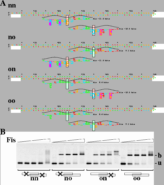
\scalebox{0.69}{\includegraphics*{fisori.ps}} A. Design of wild-type and mutated Fis sites from E. coli oriC. Four hairpin oligos were designed and designated nn, no, on and oo where n means no site because of engineered mutations (pink boxes, with information less than zero) and o means that there is a complete wild-type origin Fis site (green boxes, with positive information). For example, no contains only the Fis site closest to R3 on the right side. B. Gel mobility shift assay with oriC sites using the oligos shown in part A at a concentration of 10 nM each. Fis concentrations were 0, 30, 100, 300 and 1000 nM. u: unbound DNA; b: Fis bound DNA.


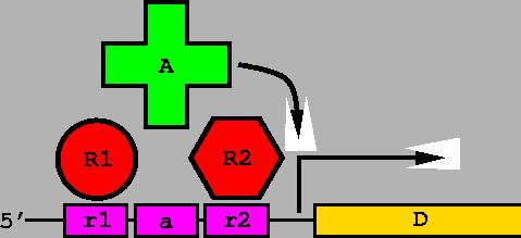
Figure 8: NOR Gate molecular computer.

An activator protein molecule A (green plus) binds to a DNA molecule at position a. When the activator binds, it turns on the promoter for gene D. Two repressor protein molecules R1 and R2 (red circle and red hexagon respectively) bind to DNA at positions r1 and r2. Binding to either r1 or r2 interferes with binding by A, so the activator can only bind when the two repressors are absent. Assigning the presence of a molecule as `1' or `true' and the absence as `0' or `false', then D = R1 NOR R2. By connecting such NOR gates together, any computer circuit can be built.

$\;$


\scalebox{1.00}{\includegraphics*{cover.ps}}
Proposed cover figure. ``Fis binding sites form a bi-stable flip-flop in the Escherichia coli origin of replication.''


next up previous
Next: About this document ... Up: Molecular Flip-Flops Formed by Previous: Acknowledgments
Tom Schneider
2003-10-16